
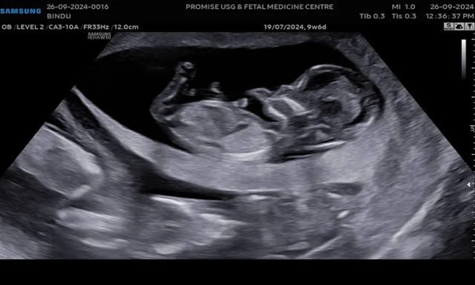
An abnormal result on the nuchal translucency (NT) scan can be worrying for expectant parents. However, it helps to know that this scan is a screening test, and not a diagnosis. An abnormal finding shows a higher chance of certain conditions, and further testing is what provides clear answers.
What Is the NT Scan?
The NT scan is an ultrasound done between 11 weeks and 13 weeks 6 days of pregnancy. It measures the small fluid-filled space at the back of the baby’s neck (called nuchal translucency). Every baby has some fluid here, but a thicker measurement can be linked to chromosomal or structural conditions.
The scan is usually combined with a blood test that measures PAPP-A and free beta-hCG. Together with the mother’s age, these results are used to estimate the chance of conditions such as Down syndrome. This approach is called combined first-trimester screening.
What Does an Abnormal Result Mean?
An NT measurement of 3.5 mm or more is commonly considered increased, although some centres use 3.0 mm or the 95th percentile for gestational age. A result may also be called abnormal when the combined screening gives a high-risk figure. The cut-off for this varies between laboratories.
A higher chance is not a confirmed condition. Many babies with increased NT are born healthy, particularly when the measurement is mildly raised, and later tests are normal.
Possible Causes
Increased NT can be associated with:
- Chromosomal conditions such as Down syndrome (trisomy 21), Edwards syndrome (trisomy 18), Patau syndrome (trisomy 13) and Turner syndrome
- Congenital heart defects
- Other structural problems, such as diaphragmatic hernia or skeletal conditions
- Rare genetic syndromes
- No identifiable cause, which is also common
Technical factors, such as the baby’s position or inaccurate dating of the pregnancy, can sometimes affect the measurement. This is why the scan should be done by a trained and accredited sonographer.
Next Steps
Your doctor will first explain the result and go through your individual risk. Follow-up usually includes:
1. Cell-free DNA test (NIPT):
A blood test that screens for common chromosomal conditions with high accuracy. It is still a screening test, not a diagnosis.
2. Diagnostic testing:
Chorionic villus sampling (CVS) can be done from about 11 to 14 weeks. Amniocentesis is usually done after 15-20 weeks. Both analyse the baby’s genetic material and give a definite answer. Each carries a small risk of miscarriage, which your doctor will discuss with you.
3. Detailed anomaly scan:
Done at around 18 to 22 weeks to examine the baby’s organs and structure closely.
4. Fetal echocardiography:
A specialised heart scan, often advised because a raised NT is linked to heart defects even when chromosomes are normal.
5. Follow-up growth scans:
These monitor the baby’s development through pregnancy.
Questions to Ask Your Doctor
- What was my exact NT measurement, and what is my risk figure?
- Which test is most suitable for me, and when should I have it?
- What are the risks and benefits of diagnostic testing?
- Should I see a fetal medicine specialist or genetic counsellor?
Final Thoughts
An abnormal NT scan ultrasound is a reason for further evaluation, not a reason to assume the worst. Timely testing, clear information and support from a qualified gynaecologist and obstetrician or fetal medicine specialist help you make informed decisions. Always rely on your own healthcare provider for advice specific to your pregnancy.
