
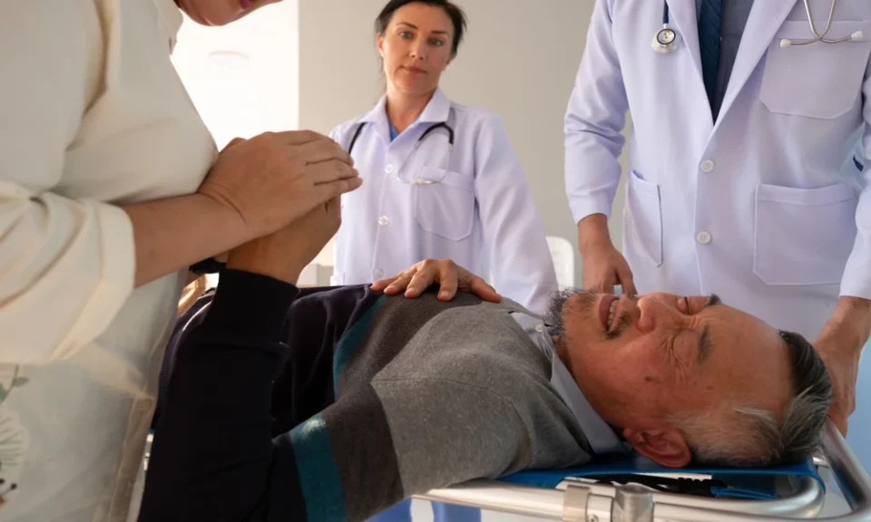
Shock is one of the most time-critical conditions a nurse will ever manage. It isn’t a single diagnosis – it’s a physiological state in which the circulatory system fails to deliver enough oxygen to the body’s tissues. Left unrecognized, even for a few minutes, shock can progress from reversible to fatal. That’s why understanding the different types of shock isn’t just test material; it’s the foundation of rapid, life-saving bedside judgment for nurses working in emergency departments, ICUs, med-surg floors, and everywhere in between.
What Does Shock Mean Physiologically?
At its core, shock is defined as inadequate tissue perfusion – the body’s cells aren’t getting enough oxygen and nutrients to function normally, and waste products aren’t being cleared efficiently. Blood pressure readings can look deceptively normal in early stages, which is exactly why so many clinicians are trained to look past the numbers and toward subtler clues – skin color and temperature, mentation, capillary refill time, and urine output. Recognizing shock early, before hypotension sets in, is what separates a good outcome from a preventable one, since compensatory mechanisms can mask a patient’s true instability for a surprisingly long time.
The 5 Major Types of Shock
Clinically, shock is grouped into five major categories, each with a distinct underlying mechanism. Hypovolemic shock results from significant fluid or blood loss – hemorrhage, severe dehydration, or extensive burns – reducing the volume of circulating blood available to perfuse organs. Cardiogenic shock occurs when the heart itself can’t pump effectively, often following a massive myocardial infarction or severe heart failure exacerbation. Distributive shock covers three subtypes with a shared feature of abnormal vasodilation: septic shock (from an overwhelming infection triggering systemic inflammation), anaphylactic shock (a severe, immune-mediated allergic reaction), and neurogenic shock (from spinal cord injury disrupting sympathetic vascular tone). Finally, obstructive shock happens when something physically blocks blood flow into or out of the heart, such as a massive pulmonary embolism, cardiac tamponade, or tension pneumothorax.
Recognizing Early vs. Late-Stage Shock Symptoms
Early, or compensated, shock often presents with tachycardia, mild anxiety or restlessness, and cool, pale extremities as the body reflexively shunts blood away from the periphery and toward vital organs like the brain and heart. As compensation begins to fail, blood pressure drops, mentation worsens, and urine output falls as the kidneys receive less perfusion. Late-stage, or decompensated, shock brings profound hypotension, multi-organ dysfunction, and a rapidly narrowing treatment window where interventions become progressively less effective. The real skill isn’t just spotting shock once it’s obvious on a monitor – it’s catching the compensatory phase before decompensation begins, often based on subtle trends rather than a single abnormal value.
How Clinicians Differentiate Shock Types at the Bedside
Bedside differentiation relies on a combination of history, vital sign trends, and physical exam findings, and getting it right matters enormously because treatments diverge sharply. A trauma patient with tachycardia and cool, clammy skin points toward hypovolemic shock. A patient with jugular venous distension, pulmonary crackles, and a recent cardiac event suggests cardiogenic shock. Fever, a known infection source, and warm flushed skin point toward early sepsis. A sudden reaction after medication administration, new food exposure, or an insect sting raises immediate suspicion for anaphylaxis. Matching the clinical picture to the underlying mechanism – rather than treating “low blood pressure” as one uniform problem – is what guides the correct, sometimes opposite, intervention.
Treatment Priorities by Shock Type
Treatment priorities diverge sharply by type, which is exactly why misclassification is dangerous. Hypovolemic shock calls for aggressive fluid or blood product resuscitation to restore circulating volume. Cardiogenic shock requires much more caution with fluids, since an already-struggling heart can be pushed into pulmonary edema, and often needs inotropic or vasopressor support instead. Septic shock demands early, aggressive antibiotics alongside fluid resuscitation per current sepsis bundle protocols. Anaphylaxis is treated immediately with intramuscular epinephrine, not antihistamines alone. Obstructive causes need the underlying obstruction physically relieved – think needle decompression for a tension pneumothorax or pericardiocentesis for tamponade. Applying fluids liberally to a cardiogenic shock patient, for instance, can actively worsen their condition rather than help it.
Why Trending Vital Signs Matters More Than a Single Reading
For nurses and other clinicians, mastering shock recognition means training yourself to trend vital signs over time rather than react to a single reading in isolation, to correlate patient history with the presenting picture, and to escalate concerns early rather than waiting for textbook hypotension to appear. This kind of pattern recognition is a core competency across emergency, critical care, and general med-surg settings alike, and it remains a frequent focus of nursing continuing education requirements for license renewal across nearly every state board.
The Role of Documentation in Shock Recognition
Documentation also plays a quietly important role in shock recognition, even though it’s easy to overlook in a fast-moving situation. Charting isolated vital signs without context makes it harder for the next clinician – whether that’s an oncoming shift nurse, a rapid response team, or a physician reviewing the chart remotely – to recognize a developing trend. Noting not just the numbers but the trajectory (“heart rate up from baseline of 78 to 112 over two hours, skin increasingly cool”) gives the full care team a much clearer picture than a single vital sign check ever could, and it’s often that documented trend, more than any single number, that triggers the right escalation at the right time.
Clinical Scenario: Spotting Shock Before It Escalates
Consider a practical scenario: a post-op patient develops tachycardia, a widening pulse pressure, and a slightly elevated temperature over several hours. On its own, tachycardia could suggest pain, anxiety, hypovolemia, or early sepsis, and the correct response depends entirely on which one is actually happening. A nurse who checks the surgical site for signs of infection, reviews recent intake and output, and correlates the trend with the patient’s baseline vitals is far more likely to catch developing distributive shock before it becomes hemodynamically unstable than one who treats a single elevated heart rate reading in isolation. This kind of layered, trend-based assessment is what separates reactive nursing from proactive nursing, and it’s exactly the skill set that structured shock education is designed to build.
Continuing Education for Shock Recognition
If you want a deeper, structured review of each shock category – complete with assessment findings, lab correlations, and intervention priorities – the Shock Types course lays it out step by step. It’s available as part of a broader catalog of online nursing ceu courses designed to fit around a working nurse’s schedule, whether you’re renewing this cycle or simply want a sharper, more confident recall of shock presentations the next time a patient starts to trend the wrong direction.
